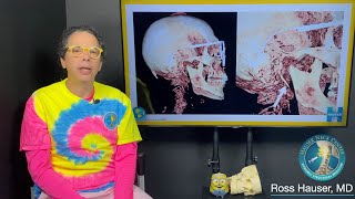
At a Glance: Micro-computed tomography (microCT) or X-Ray Microscopy (XRM) is X-ray imaging in 3D, the same method used in clinical CT ...

Show Soft Tissue On Cbct Scan - System Summary

Technical Overview

Overview for Show Soft Tissue On Cbct Scan.

Integration Notes

Authentication Context related to Show Soft Tissue On Cbct Scan.

Directory Details

Directory Access Notes about Show Soft Tissue On Cbct Scan.

What to Check First

Implementation Considerations for this topic.

Important details found

  • Micro-computed tomography (microCT) or X-Ray Microscopy (XRM) is X-ray imaging in 3D, the same method used in clinical CT ...

Why this topic is useful

A structured page helps reduce disconnected snippets by grouping the main subject with context, examples, and nearby entries.

Sponsored

What to Check First

What does Show Soft Tissue On Cbct Scan usually refer to?

Show Soft Tissue On Cbct Scan usually relates to authentication, directory access, identity handling, or system integration context within a technical environment.

Can this information vary between systems?

Yes. LDAP, SSO, directory access, and identity configurations can vary by provider, software version, and enterprise policy.

What does Show Soft Tissue On Cbct Scan usually refer to?

Show Soft Tissue On Cbct Scan usually relates to authentication, directory access, identity handling, or system integration context within a technical environment.

Visual References

Show soft tissue on CBCT scan
Visualizing soft tissue in Cone-Beam CT
Unknown soft tissue calcification on CBCT
Soft Tissue & Bone - 3D Cone Beam CT Scan and Animation by SmartScan Imaging of Orange
Simple Workflows for Challenging Cases: The Role of Soft Tissues During CBCT Scanning
Cone beam CT scan showing elongated styloids, misalignments, and more. Hauser Neck Center
Soft Tissue & MicroCT | Applications of MicroCT in Life Science #6
Sponsored
View Full Details
Show soft tissue on CBCT scan

Show soft tissue on CBCT scan

Read more details and related context about Show soft tissue on CBCT scan.

Visualizing soft tissue in Cone-Beam CT

Visualizing soft tissue in Cone-Beam CT

The use of Contrax3D can visualize the mucosa. This means that surgical guides can be manufactured on the bases of

Unknown soft tissue calcification on CBCT

Unknown soft tissue calcification on CBCT

Provide your differential diagnosis in the comment section. Faculty webpage

Soft Tissue & Bone - 3D Cone Beam CT Scan and Animation by SmartScan Imaging of Orange

Soft Tissue & Bone - 3D Cone Beam CT Scan and Animation by SmartScan Imaging of Orange

Read more details and related context about Soft Tissue & Bone - 3D Cone Beam CT Scan and Animation by SmartScan Imaging of Orange.

Simple Workflows for Challenging Cases: The Role of Soft Tissues During CBCT Scanning

Simple Workflows for Challenging Cases: The Role of Soft Tissues During CBCT Scanning

Read more details and related context about Simple Workflows for Challenging Cases: The Role of Soft Tissues During CBCT Scanning.

Cone beam CT scan showing elongated styloids, misalignments, and more. Hauser Neck Center

Cone beam CT scan showing elongated styloids, misalignments, and more. Hauser Neck Center

Read more details and related context about Cone beam CT scan showing elongated styloids, misalignments, and more. Hauser Neck Center.

Soft Tissue & MicroCT | Applications of MicroCT in Life Science #6

Soft Tissue & MicroCT | Applications of MicroCT in Life Science #6

Micro-computed tomography (microCT) or X-Ray Microscopy (XRM) is X-ray imaging in 3D, the same method used in clinical CT ...